IET Prestige Lecture: Advanced materials and 3D printing for the regeneration of human tissues
Presented by Professor Kevin Shakesheff
Churchill College, Cambridge, February 2015
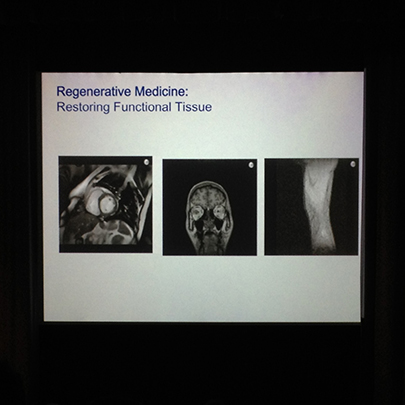
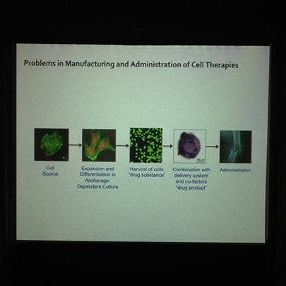
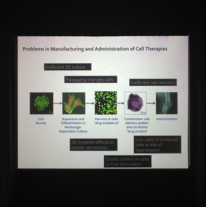
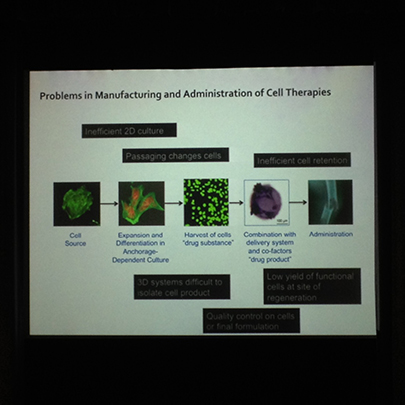
The focus of the talk was the still-emerging technology and techniques of 3D printing for the purpose of regenerating human tissue, and the challenges faced in their use and development. The challenges were specified by Professor Shakesheff during his talk as;
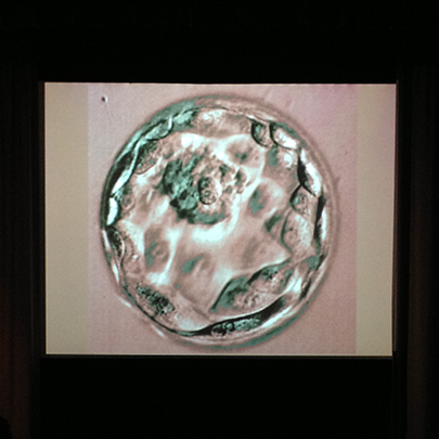
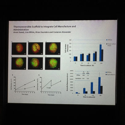
Growing stem cells into the correct tissue
Stem cells are used for growing the required new tissue but the nature of these means that they can grow to form any type of cells within the body. The challenge with these is therefore how to control their growth so that they grown into the correct type of cells for the required type of tissue or body part. Even if this challenge is overcome, a further challenge is to grow the cells in three dimensions, as is usually required for the body part. This is usually not possible in a typical dish in the lab — two dimensional growth is the best achievable in this setting.
Delivery
In some cases cells may be simply injected into the body in the required location and will form new cells of the the correct type due to the surrounding cells. In other cases delivery is more complex and requires patterning of cells grown outside the body to create the required component before delivery.
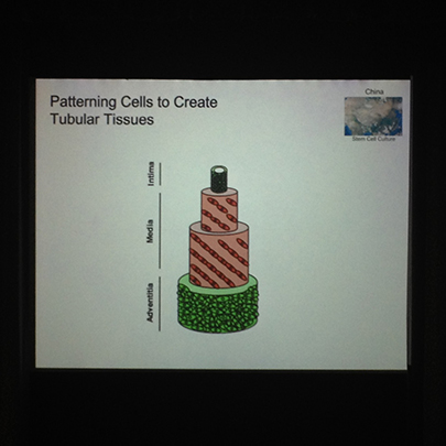
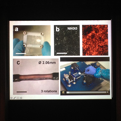
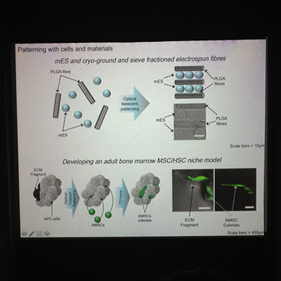
Patterning
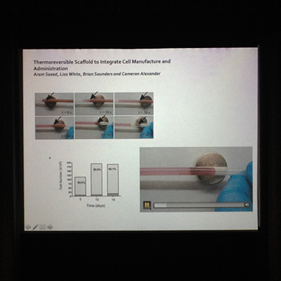
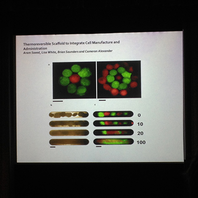
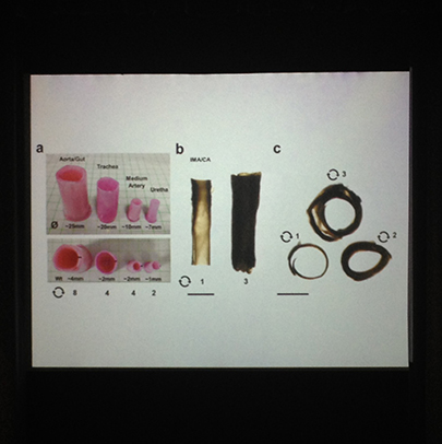
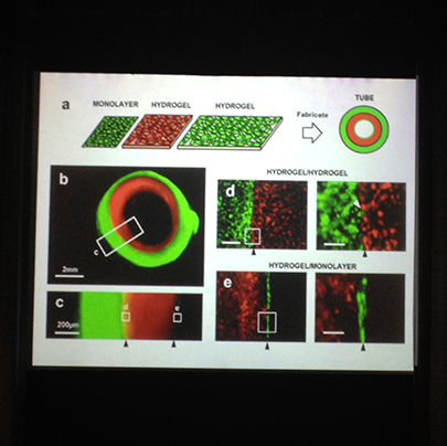
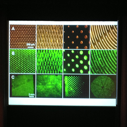
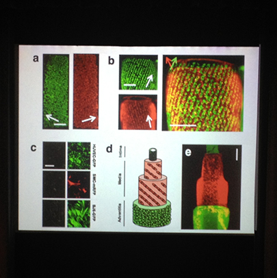
Patterning — i.e shaping the cells to form the required shape of a biological component is an ongoing challenge. Some innovative methods for creating tubular tissue include rolling up the tissue once grown as a flat piece and growing the tissue around a tubular mould. These tubes require three layers of different tissue. A biodegradable scaffold is often used, which is placed within the body at the required location — i.e. a scaffold the shape of a nose — and the new tissue is grown on the scaffold in situ.
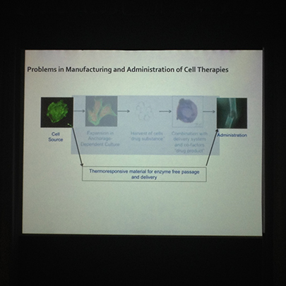
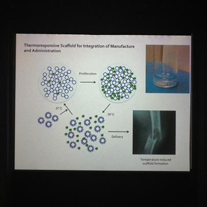
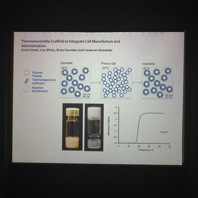
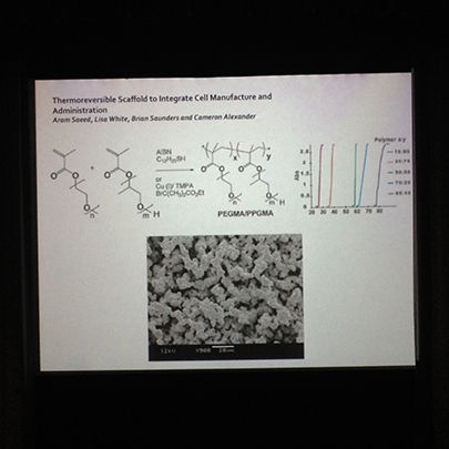
Biodegradable scaffold material
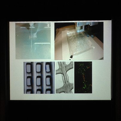
Professor Shakesheff discussed the chemistry of the scaffold material on which the new cells are grown, which has been developed with the required biodegradable properties as well as to be compatible with human tissue.
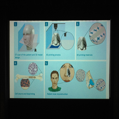
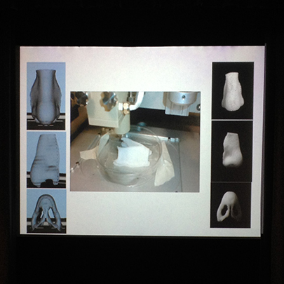
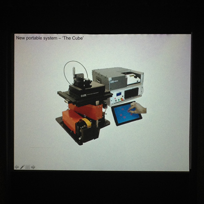
Scanning and printing with the biodegradable scaffold material
The scanning and printing process was described. This involves first taking a CT scan and 3D mould of the body part or area to be reconstructed, along with the surrounding area. There may be the opportunity to do this before removal of the body part due to the discovery of a cancerous growth, for example. The next stage is the 3D printing process, the material which is printed being the biodegradable material that will form the bio scaffolding for the new body part. The shape to which this is printed is informed by the CT scan and mould. The next stage is sourcing the cells that are to be bioprinted onto the bio scaffold material. These cells are usually harvested from another part of the patients body or from a donor source, a typical example being bone marrow cells. The bio scaffold material along with the new cells are then surgically placed into the patient's body where the new cells grow to join existing surround tissue.
Conclusion
Much work is still to be done in developing these techniques, but the hope is that in the medium term 3D printing for the generation of living tissue will be able to replace the complex work of plastic surgeons.